
Clinical case of an extruded lumbar herniated disc
Published in BACKspace, the leading magazine of the ECU (European Chiropractic Union).
On 11/02/06, Mrs. Elisabetta, 42 years old, Caucasian, came to my clinic accompanied by her husband because she was unable to stand due to severe back pain. She began telling me her story, stating that chiropractic was her last hope.
This is her story.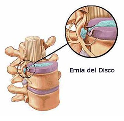
She said she had not slept for three nights because of unbearable back pain radiating down her right leg, from the buttock to the foot, and that she could not find any position that gave her relief. She reported having suffered from back pain since she was young and having already been examined and treated by several spine specialists, but she had never consulted a chiropractor.
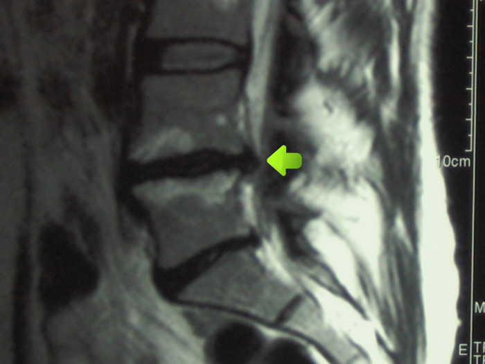
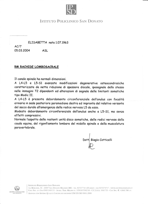
In 2004, a neurosurgeon proposed surgery after examining her and reviewing the MRI result (Fig. 1), which showed a clear herniated disc between L4 and L5. However, she refused the operation, preferring drug therapy with injections of Voltaren and Muscoril.
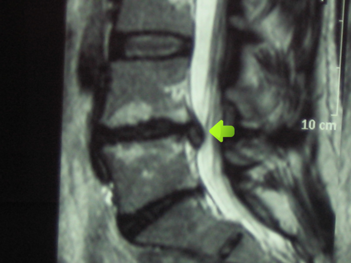
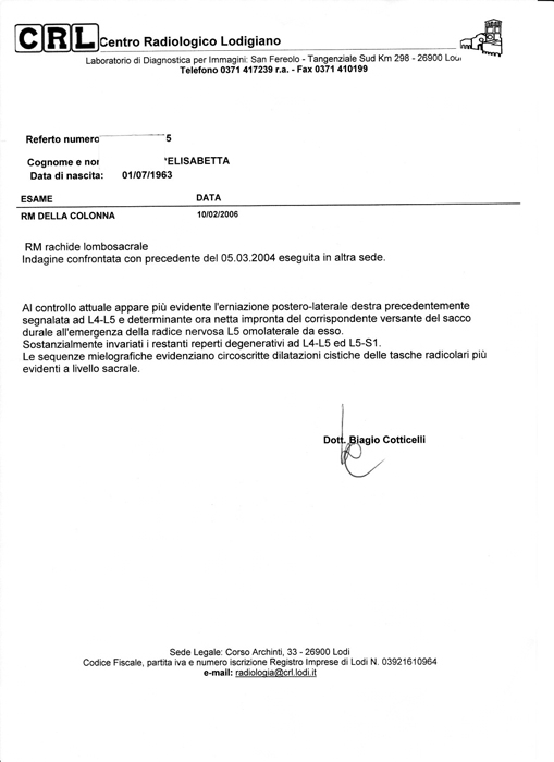
After the drug therapy, the pain disappeared until January 2006, when one morning she began to feel a stabbing pain in the lumbar region radiating down her right leg. Her doctor prescribed the same drug therapies that had helped her in the past, but the pain did not lessen. Her general practitioner then decided to replace anti-inflammatories with a corticosteroid treatment, which provided no symptom relief. For this reason, another MRI (Fig. 2) was requested, showing a clear worsening of the same disc herniation with compression of the right L5 nerve root. At that point, she was advised to see a neurosurgical specialist.
After the consultation, the neurosurgeon again suggested an urgent surgical operation, but the patient refused surgery once more.
On the advice of a family friend, she decided to consult a chiropractor as the last possible treatment before surgery.
During the chiropractic examination, I noted a radiculopathy along the entire L5 dermatome on the right leg. The Lasègue test was positive at 20° on the right leg, where it began to cause back pain referred into the right leg. The right big toe extensor muscle was very weak. In addition, the patient tested positive on kinesiological tests used to identify dura mater tension.
I began the first cycle of chiropractic sessions: two sessions per week for four weeks, treating the patient with adjustments of vertebral subluxations along the entire spine.
After one month, the patient was asymptomatic; neurological and orthopedic tests were negative, so I recommended exercises to keep the spine mobile and advised monthly treatment for six months.
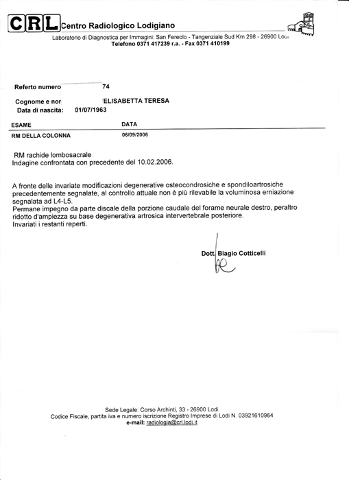
In September 2006, the patient underwent the last MRI (Fig. 3), which showed that the disc herniation had disappeared. The patient felt well, so I recommended a chiropractic check-up every six months, in addition to continuing physical exercises.
Initial phase
A clear herniated disc between L4 and L5 (05/03/2004)
MRI image
Report
Second phase
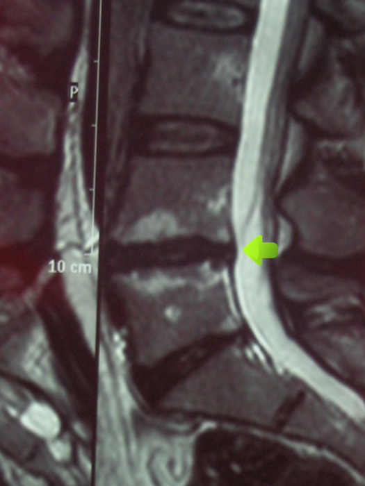
The L4–L5 disc herniation is more evident compared to 2004; the patient begins chiropractic treatments (10/02/2006)
MRI image
Report
Third phase
Six months after starting chiropractic treatments, the herniated disc between L4 and L5 has disappeared (06/09/2006)
MRI image
Report
“THE POWER THAT MAKES THE BODY HEALS THE BODY” D.D. PALMER
“La forza che crea il corpo umano lo guarisce” – D.D. Palmer (fondatore della chiropratica 1895)
Book your visit
To book an appointment at our Milan office:
Tel: +39 02 89.40.49.90
WhatsApp: +39 393 81.04.359
Email: segreteria@studiomazzinisrl.it
Online consultation
If you live outside Milan, you can book your consultation online.
Progression of complete healing of the herniated disc.
First phase
Second phase
Third phase
The images show how the herniated disc between L4 and L5, indicated by the arrow, which had worsened over time without adequate care, then completely healed after the patient received chiropractic care.